Competition between Polar and Nonpolar Lattice Distortions in Oxide Quantum Wells: New Critical Thickness at Polar Interfaces
Competition between Polar and Nonpolar Lattice Distortions in Oxide Quantum Wells: New Critical Thickness at Polar Interfaces
J. Gazquez, M. Stengel, R. Mishra, M. Scigaj, M. Varela, M. A. Roldan, J. Fontcuberta, F. Sánchez, and G. Herranz. Phys. Rev. Lett. 119, 106102 – Published 7 September 2017. DOI: https://doi.org/10.1103/PhysRevLett.119.106102
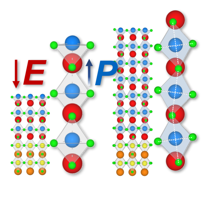
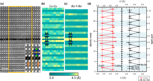
Two basic lattice distortions permeate the structural phase diagram of oxide perovskites: antiferrodistortive (AFD) rotations and tilts of the oxygen octahedral network and polar ferroelectric modes. With some notable exceptions, these two order parameters rarely coexist in a bulk crystal, and understanding their competition is a lively area of active research. Here we demonstrate, by using the LaAlO3/SrTiO3 system as a test case, that quantum confinement can be a viable tool to shift the balance between AFD and polar modes and selectively stabilize one of the two phases. By combining scanning transmission electron microscopy (STEM) and first-principles-based models, we find a crossover between a bulklike LaAlO3 structure where AFD rotations prevail, to a strongly polar state with no AFD tilts at a thickness of approximately three unit cells; therefore, in addition to the celebrated electronic reconstruction, our work unveils a second critical thickness, related not to the electronic properties but to the structural ones. We discuss the implications of these findings, both for the specifics of the LaAlO3/SrTiO3 system and for the general quest towards nanoscale control of material properties.


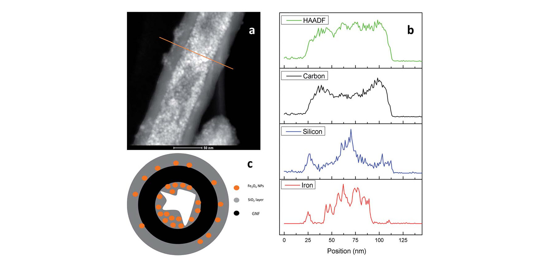
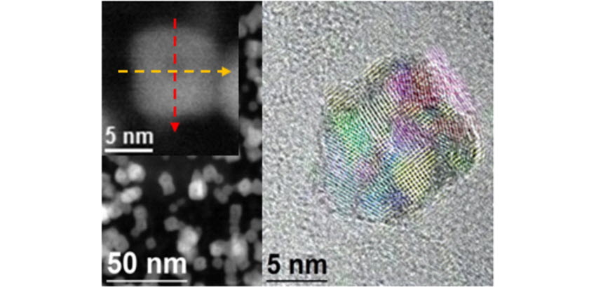
 relaxivities for Fe3O4@GNF nanocomposites and Fe3O4@GNF@SiO>2 nanocapsules revealing the potential application of these hybrid materials for bioimaging. Therefore, the coating of filled GNF with silica is as an excellent strategy for the protection of encapsulated payloads.
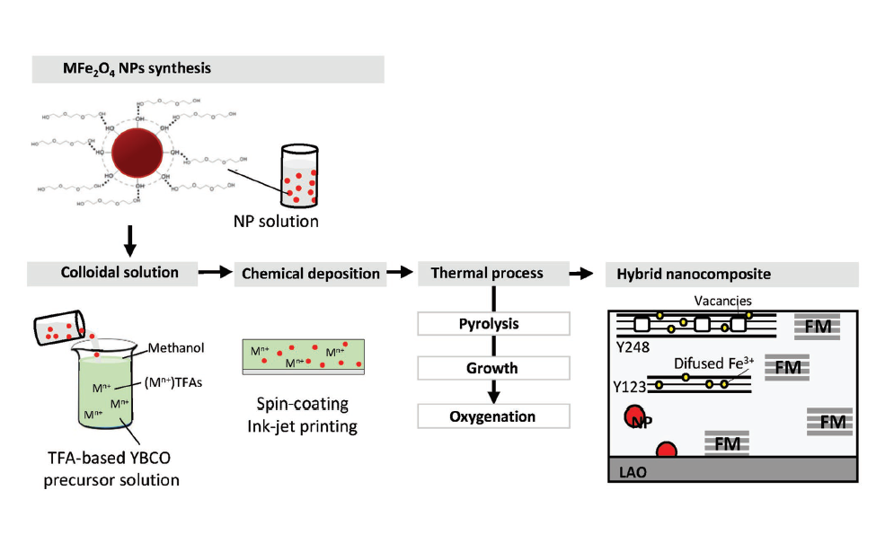


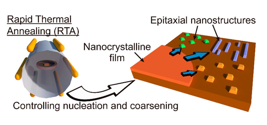

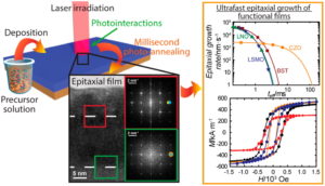
 Katrien De Keukeleere, Pablo Cayado, Alexander Meledin, Ferran Vallès, Jonathan De Roo, Hannes Rijckaert, Glenn Pollefeyt, Els Bruneel, Anna Palau, Mariona Coll, Susagna Ricart, Gustaaf Van Tendeloo, Teresa Puig, Xavier Obradors, Isabel Van Driessche. Advanced Electronic Materials.